 ICU Basics
Cheat Sheet
TEG and ROTEM: Reading Viscoelastic Tests
What the tracing shows that PT/INR and aPTT miss, each value and the product it points to, example ranges, and TXA timing
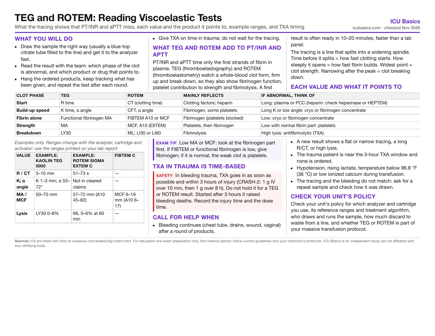
What you will do
- Draw the sample the right way (usually a blue-top citrate tube filled to the line) and get it to the analyzer fast.
- Read the result with the team: which phase of the clot is abnormal, and which product or drug that points to.
- Hang the ordered products, keep tracking what has been given, and repeat the test after each round.
- Give TXA on time in trauma; do not wait for the tracing.
What TEG and ROTEM add to PT/INR and aPTT
PT/INR and aPTT time only the first strands of fibrin in plasma. TEG (thromboelastography) and ROTEM (thromboelastometry) watch a whole-blood clot form, firm up and break down, so they also show fibrinogen function, platelet contribution to strength and fibrinolysis. A first result is often ready in 10–20 minutes, faster than a lab panel.
The tracing is a line that splits into a widening spindle. Time before it splits = how fast clotting starts. How steeply it opens = how fast fibrin builds. Widest point = clot strength. Narrowing after the peak = clot breaking down.
Each value and what it points to
| Clot phase | TEG | ROTEM | Mainly reflects | If abnormal, think of |
|---|---|---|---|---|
| Start | R time | CT (clotting time) | Clotting factors; heparin | Long: plasma or PCC (heparin: check heparinase or HEPTEM) |
| Build-up speed | K time, α angle | CFT, α angle | Fibrinogen, some platelets | Long K or low angle: cryo or fibrinogen concentrate |
| Fibrin alone | Functional fibrinogen MA | FIBTEM A10 or MCF | Fibrinogen (platelets blocked) | Low: cryo or fibrinogen concentrate |
| Strength | MA | MCF, A10 (EXTEM) | Platelets, then fibrinogen | Low with normal fibrin part: platelets |
| Breakdown | LY30 | ML; LI30 or LI60 | Fibrinolysis | High lysis: antifibrinolytic (TXA) |
| Value | Example: kaolin TEG 5000 | Example: ROTEM sigma EXTEM C | FIBTEM C |
|---|---|---|---|
| R / CT | 5–10 min | 51–73 s | — |
| K; α angle | K 1–3 min; α 53–72° | Not in cleared claims | — |
| MA / MCF | 50–70 mm | 57–72 mm (A10 45–62) | MCF 6–19 mm (A10 6–17) |
| Lysis | LY30 0–8% | ML 0–6% at 60 min | — |
TXA in trauma is time-based
Call for help when
- Bleeding continues (chest tube, drains, wound, vaginal) after a round of products.
- A new result shows a flat or narrow tracing, a long R/CT, or high lysis.
- The trauma patient is near the 3-hour TXA window and none is ordered.
- Hypotension, rising lactate, temperature below 96.8 °F (36 °C) or low ionized calcium during transfusion.
- The tracing and the bleeding do not match: ask for a repeat sample and check how it was drawn.
Check your unit's policy
Check your unit's policy for which analyzer and cartridge you use, its reference ranges and treatment algorithm, who draws and runs the sample, how much discard to waste from a line, and whether TEG or ROTEM is part of your massive transfusion protocol.
In depth
The one-page summary above is what the PDF prints. These sections go further.
How the test works: analyzers and assays
Both systems put a small whole-blood sample at 37 °C, add calcium and an activator, and measure how stiff the clot becomes over time. Amplitude is in millimeters; time runs left to right. TEG 5000 uses a cup and pin; TEG 6s and ROTEM sigma use closed cartridges that run several assays at once.
| System | Assay | What it does |
|---|---|---|
| TEG | Kaolin (CK) | Contact activation; R, K, angle, MA, LY30 |
| TEG | RapidTEG (CRT) | Tissue factor plus kaolin; fast MA (and TEG-ACT on TEG 5000) |
| TEG | Functional fibrinogen (CFF) | Platelets blocked; MA shows fibrin strength |
| TEG | Kaolin with heparinase (CKH) | Heparin removed; compare R with plain kaolin |
| ROTEM | EXTEM | Tissue factor (extrinsic) activation |
| ROTEM | INTEM | Contact (intrinsic) activation; sensitive to heparin |
| ROTEM | FIBTEM | EXTEM with platelets blocked: fibrin part only |
| ROTEM | HEPTEM | INTEM with heparinase: shows heparin effect |
| ROTEM | APTEM (delta) | EXTEM with an antifibrinolytic: confirms lysis |
Definitions: R and CT run from the start to a 2 mm amplitude. K and CFT run from 2 to 20 mm. The α angle is the slope of the opening curve. MA and MCF are the widest amplitude; A5, A10 and A20 are the amplitude 5, 10 and 20 minutes after CT, so A10 gives an early read of final strength. LY30 is the percent loss of clot 30 minutes after MA; ROTEM LI30 or LI60 is the percent of clot still left (a low number means lysis), and ML is the maximum percent lost.
The FDA-cleared TEG 6s trauma cartridge reports only CK R and LY30, CRT MA and CFF MA, with no K or angle. On that device, read fibrinogen from the functional fibrinogen MA. The US ROTEM sigma cartridge reports CT, A5 to A20, MCF, LI60 and ML.
Reading common patterns
No drawn tracing yet: picture a thin line that opens into a spindle. These are the shapes to recognize.
| Shape | Values | Likely cause | Usual response |
|---|---|---|---|
| Long straight line before it opens, then normal width | Long R or CT | Factor deficiency, dilution, warfarin; heparin if heparinase/HEPTEM corrects it | Plasma or PCC; protamine if heparin |
| Opens slowly, stays narrow | Long K, low angle, low FIBTEM or functional fibrinogen | Low fibrinogen (dilution, consumption, obstetric bleeding) | Cryo or fibrinogen concentrate |
| Opens on time but stays narrow | Low MA or MCF, normal FIBTEM | Low platelet count or function | Platelets |
| Opens, then pinches closed early | High LY30 or ML, low LI30/LI60 | Hyperfibrinolysis (trauma, liver surgery) | Antifibrinolytic |
| Opens fast and very wide | Short R, high MA | Hypercoagulable state | Discuss VTE prophylaxis; no products |
Example treatment triggers from published algorithms
Programs set their own cutoffs. These are the ACS TQIP 2014 massive transfusion examples, given here to show the logic; your hospital may use different numbers (the Blood Products page lists a Denver rapid-TEG set and a European ROTEM set).
| Finding | Standard TEG | Rapid TEG | ROTEM | Product |
|---|---|---|---|---|
| Slow start | R >9 min | ACT >128 s | EXTEM CT >100 s or INTEM CT >230 s | Plasma |
| Slow build-up | K >4 min; α <60° | K >2.5 min; α <60° | FIBTEM MCF <8 mm | Cryo or fibrinogen concentrate and/or plasma |
| Weak clot | MA <55 mm | MA <55 mm | EXTEM MCF <45 mm with FIBTEM MCF >10 mm | Platelets |
| Lysis | LY30 >7.5% | LY30 >3% | EXTEM ML >15% | Antifibrinolytic |
The European trauma guideline (6th ed., 2023) recommends a goal-directed strategy using standard labs and/or viscoelastic tests, fibrinogen concentrate or cryo (starting 3–4 g) for a viscoelastic fibrin deficit or Clauss fibrinogen 150 mg/dL (1.5 g/L) or less, avoiding plasma to correct low fibrinogen when cryo or concentrate is available, and PCC when initiation stays slow with normal fibrinogen. Ionized calcium, temperature and pH still need correcting; the test cannot show them.
Sample handling and what the test misses
- Use the tube your lab specifies (usually 3.2% sodium citrate). Fill to the line: an underfilled tube has too much citrate.
- Avoid heparin contamination from a flushed arterial or central line: waste the discard volume your policy sets, or draw from another site. A long R that corrects with heparinase or HEPTEM points to heparin.
- Label with the draw time and send right away; follow the lab's time limit for running citrated samples.
- The test runs at 37 °C, so it hides the effect of hypothermia. Warm the patient anyway.
- The sample is recalcified, so it hides low ionized calcium. Check an ionized calcium separately.
- Standard TEG and ROTEM do not detect aspirin or P2Y12 inhibitors, and they do not reliably show DOAC or low-molecular-weight heparin effect. Platelet mapping or drug levels are separate tests.
Where it is used and the evidence
- Trauma: guides products after the first ratio-based MTP rounds. Gonzalez 2016 (single center, 111 patients) found lower 28-day mortality with a TEG-guided MTP (19.6% vs 36.4%) and less early plasma and platelet use. The multicenter iTACTIC trial (396 patients, 2021) found no difference when both arms started from empiric ratios.
- Cardiac surgery: the largest share of trials. Bleeding after bypass, with heparin, protamine and a pump-depleted platelet pool, is where a fast read of R, fibrinogen and MA helps most.
- Liver transplant and liver surgery: INR overstates bleeding risk in liver disease, and hyperfibrinolysis is common during reperfusion.
- Obstetric hemorrhage: fibrinogen falls early, and FIBTEM or functional fibrinogen gives a quick fibrinogen read. TXA in postpartum hemorrhage is also time-based: the WOMAN trial cut bleeding deaths, most clearly when given within 3 hours of birth.
The Cochrane review updated in May 2026 (35 randomized trials, 3,096 patients, mostly elective cardiac surgery) found that TEG- or ROTEM-guided algorithms may lower mortality (RR 0.76) and plasma and platelet use, but the certainty of the evidence is very low.
Tranexamic acid timing in trauma
CRASH-2 (Lancet 2010) randomized 20,211 bleeding trauma patients within 8 hours of injury to TXA (1 g over 10 minutes, then 1 g over 8 hours) or placebo. All-cause death fell from 16.0% to 14.5% and bleeding death from 5.7% to 4.9%. The timing analysis (2011) found the benefit largest within 1 hour, still present at 1–3 hours, and harm after 3 hours (bleeding death 4.4% vs 3.1%).
The European guideline (2023) recommends TXA as soon as possible within 3 hours (Grade 1A) and recommends not waiting for viscoelastic results (Grade 1B). A later high LY30 can prompt more antifibrinolytic if the team decides, but the first dose is driven by the clock. Hand over the injury time, dose time and whether the 8-hour infusion is running so no one gives a duplicate bolus.
Nursing care during a viscoelastic-guided resuscitation
- Keep a running list of every product, volume and time; the next result is read against what was given.
- Repeat the test after each targeted round, usually with a CBC, fibrinogen, ionized calcium and blood gas, per protocol.
- Warm all fluids and products and the patient; replace calcium per protocol.
- Watch the drains and dressings and record output, so the team can tell surgical from coagulopathic bleeding.
- After bleeding stops, a hypercoagulable trace is common; ask about starting VTE prophylaxis.
Related cheat sheets
- Sepsis and Septic Shock BundleSepsis-3 definitions, the Hour-1 bundle and SSC 2026 changes for exam review
- Preventing ICU InfectionsCLABSI, CAUTI, VAE, C. difficile and MDRO bundles, isolation by organism, hand hygiene and quality measures
- Acute Kidney InjuryKDIGO staging, causes, urine studies, nephrotoxins, prevention, dialysis timing and nursing care for critical care exams
- Electrolyte ImbalancesNa, K, Ca, Mg and phos: normals, ECG clues, safe replacement, hyperkalemia steps
- CRRT (Continuous Renal Replacement)Modes, dose, circuit pressures, citrate and nursing care for critical care exams
- DKA and HHS2024 consensus criteria, treatment order, potassium, euglycemic DKA and monitoring
Sources
- American College of Surgeons Trauma Quality Improvement Program. ACS TQIP Massive Transfusion in Trauma Guidelines. October 2014. Link
- Rossaint R, Afshari A, Bouillon B, et al. The European guideline on management of major bleeding and coagulopathy following trauma: sixth edition. Crit Care. 2023;27(1):80. Link
- Gonzalez E, Moore EE, Moore HB, et al. Goal-directed hemostatic resuscitation of trauma-induced coagulopathy: a pragmatic randomized clinical trial comparing a viscoelastic assay to conventional coagulation assays. Ann Surg. 2016;263(6):1051-1059. Link
- Baksaas-Aasen K, Gall LS, Stensballe J, et al. Viscoelastic haemostatic assay augmented protocols for major trauma haemorrhage (ITACTIC): a randomized, controlled trial. Intensive Care Med. 2021;47(1):49-59. Link
- CRASH-2 trial collaborators. Effects of tranexamic acid on death, vascular occlusive events, and blood transfusion in trauma patients with significant haemorrhage (CRASH-2): a randomised, placebo-controlled trial. Lancet. 2010;376(9734):23-32. Link
- CRASH-2 collaborators. The importance of early treatment with tranexamic acid in bleeding trauma patients: an exploratory analysis of the CRASH-2 randomised controlled trial. Lancet. 2011;377(9771):1096-1101. Link
- WOMAN Trial Collaborators. Effect of early tranexamic acid administration on mortality, hysterectomy, and other morbidities in women with post-partum haemorrhage (WOMAN). Lancet. 2017;389(10084):2105-2116. Link
- Kvisselgaard AD, Wolthers SA, Wikkelsø AJ, et al. Thromboelastography (TEG) or thromboelastometry (ROTEM) to monitor haemostatic treatment versus usual care in adults or children with bleeding. Cochrane Database Syst Rev. 2026;5:CD007871. Link
- US Food and Drug Administration. 510(k) decision summary K201440: ROTEM sigma Thromboelastometry System (Tem Innovations / Werfen), reference intervals. 2022. Link
- US Food and Drug Administration. 510(k) decision summary K183160: TEG 6s Hemostasis System, Citrated: K, RT, FF Assay Cartridge (Haemonetics). Link