 ICU Basics
Cheat Sheet
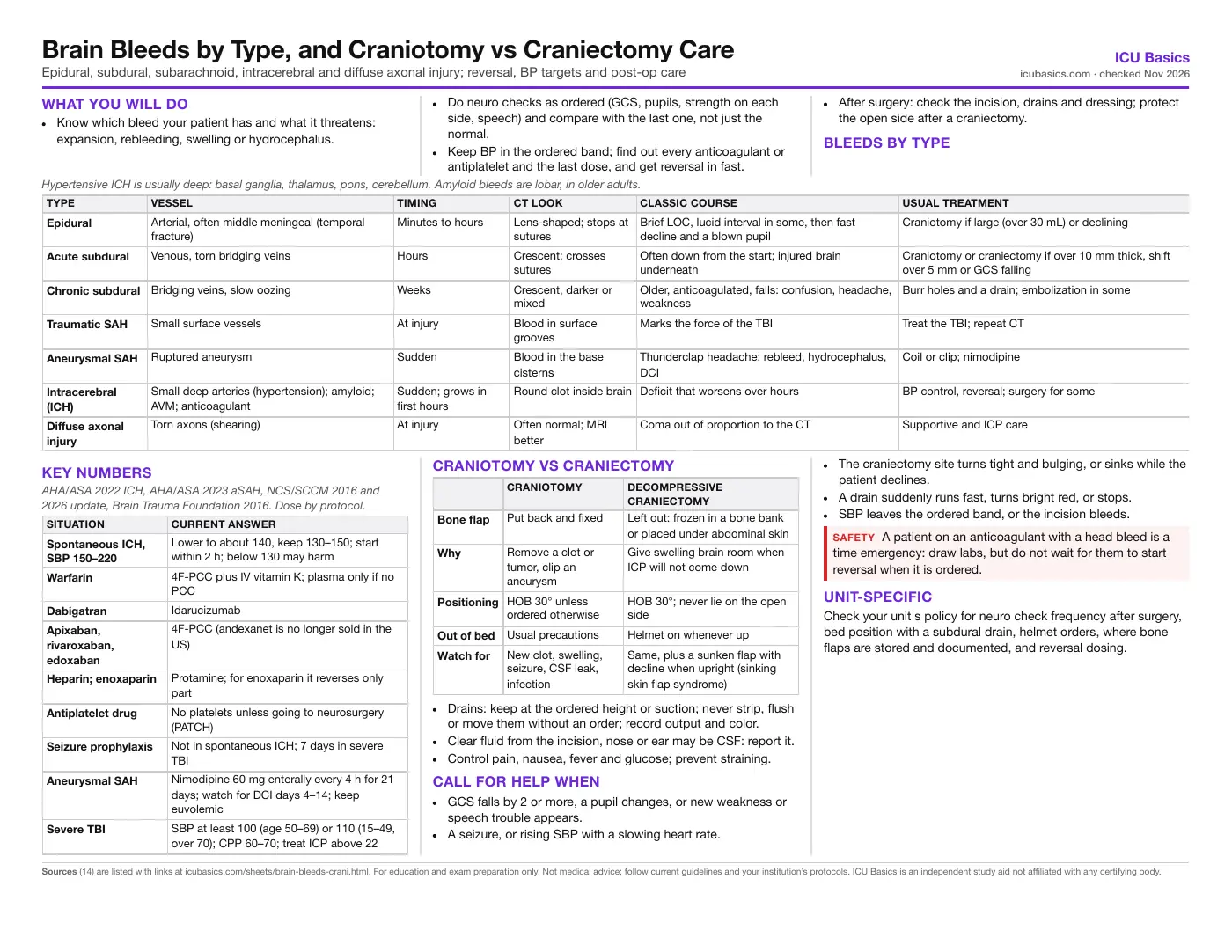
Brain Bleeds by Type, and Craniotomy vs Craniectomy Care
Epidural, subdural, subarachnoid, intracerebral and diffuse axonal injury; reversal, BP targets and post-op care
What you will do
- Know which bleed your patient has and what it threatens: expansion, rebleeding, swelling or hydrocephalus.
- Do neuro checks as ordered (GCS, pupils, strength on each side, speech) and compare with the last one, not just the normal.
- Keep BP in the ordered band; find out every anticoagulant or antiplatelet and the last dose, and get reversal in fast.
- After surgery: check the incision, drains and dressing; protect the open side after a craniectomy.
Bleeds by type
| Type | Vessel | Timing | CT look | Classic course | Usual treatment |
|---|---|---|---|---|---|
| Epidural | Arterial, often middle meningeal (temporal fracture) | Minutes to hours | Lens-shaped; stops at sutures | Brief LOC, lucid interval in some, then fast decline and a blown pupil | Craniotomy if large (over 30 mL) or declining |
| Acute subdural | Venous, torn bridging veins | Hours | Crescent; crosses sutures | Often down from the start; injured brain underneath | Craniotomy or craniectomy if over 10 mm thick, shift over 5 mm or GCS falling |
| Chronic subdural | Bridging veins, slow oozing | Weeks | Crescent, darker or mixed | Older, anticoagulated, falls: confusion, headache, weakness | Burr holes and a drain; embolization in some |
| Traumatic SAH | Small surface vessels | At injury | Blood in surface grooves | Marks the force of the TBI | Treat the TBI; repeat CT |
| Aneurysmal SAH | Ruptured aneurysm | Sudden | Blood in the base cisterns | Thunderclap headache; rebleed, hydrocephalus, DCI | Coil or clip; nimodipine |
| Intracerebral (ICH) | Small deep arteries (hypertension); amyloid; AVM; anticoagulant | Sudden; grows in first hours | Round clot inside brain | Deficit that worsens over hours | BP control, reversal; surgery for some |
| Diffuse axonal injury | Torn axons (shearing) | At injury | Often normal; MRI better | Coma out of proportion to the CT | Supportive and ICP care |
Key numbers
| Situation | Current answer |
|---|---|
| Spontaneous ICH, SBP 150–220 | Lower to about 140, keep 130–150; start within 2 h; below 130 may harm |
| Warfarin | 4F-PCC plus IV vitamin K; plasma only if no PCC |
| Dabigatran | Idarucizumab |
| Apixaban, rivaroxaban, edoxaban | 4F-PCC (andexanet is no longer sold in the US) |
| Heparin; enoxaparin | Protamine; for enoxaparin it reverses only part |
| Antiplatelet drug | No platelets unless going to neurosurgery (PATCH) |
| Seizure prophylaxis | Not in spontaneous ICH; 7 days in severe TBI |
| Aneurysmal SAH | Nimodipine 60 mg enterally every 4 h for 21 days; watch for DCI days 4–14; keep euvolemic |
| Severe TBI | SBP at least 100 (age 50–69) or 110 (15–49, over 70); CPP 60–70; treat ICP above 22 |
Craniotomy vs craniectomy
| Craniotomy | Decompressive craniectomy | |
|---|---|---|
| Bone flap | Put back and fixed | Left out: frozen in a bone bank or placed under abdominal skin |
| Why | Remove a clot or tumor, clip an aneurysm | Give swelling brain room when ICP will not come down |
| Positioning | HOB 30° unless ordered otherwise | HOB 30°; never lie on the open side |
| Out of bed | Usual precautions | Helmet on whenever up |
| Watch for | New clot, swelling, seizure, CSF leak, infection | Same, plus a sunken flap with decline when upright (sinking skin flap syndrome) |
- Drains: keep at the ordered height or suction; never strip, flush or move them without an order; record output and color.
- Clear fluid from the incision, nose or ear may be CSF: report it.
- Control pain, nausea, fever and glucose; prevent straining.
Call for help when
- GCS falls by 2 or more, a pupil changes, or new weakness or speech trouble appears.
- A seizure, or rising SBP with a slowing heart rate.
- The craniectomy site turns tight and bulging, or sinks while the patient declines.
- A drain suddenly runs fast, turns bright red, or stops.
- SBP leaves the ordered band, or the incision bleeds.
Unit-specific
Check your unit's policy for neuro check frequency after surgery, bed position with a subdural drain, helmet orders, where bone flaps are stored and documented, and reversal dosing.
In depth
The one-page summary above is what the PDF prints. These sections go further.
Epidural and subdural hematomas
An epidural hematoma sits between the skull and the dura. Most come from an artery, usually the middle meningeal artery torn by a temporal bone fracture, so they grow fast. The dura is stuck to the skull at the sutures, which is why the clot is lens-shaped and stops at the suture lines. Only some patients show the classic lucid interval (knocked out, wakes up, then declines); many are never lucid. With early surgery the outcome is often good, so speed matters.
A subdural hematoma sits under the dura, along the brain surface, from torn bridging veins. It spreads in a crescent and crosses suture lines. Acute subdurals follow high-energy injury and usually come with injured brain underneath, so outcome is worse than with an epidural. Brain shrinkage with age stretches the bridging veins, and anticoagulants, antiplatelets and alcohol add risk, so older adults can bleed after a minor fall.
| Lesion | Surgery regardless of GCS | Brain Trauma Foundation surgical guideline (2006) |
|---|---|---|
| Epidural | Over 30 mL | Under 30 mL, under 15 mm thick, shift under 5 mm and GCS over 8 without focal deficit: may be watched with serial CT |
| Acute subdural | Over 10 mm thick or midline shift over 5 mm | Comatose with a smaller clot: surgery if GCS falls by 2, pupils become unequal or fixed, or ICP exceeds 20 |
A chronic subdural develops over weeks: the patient or family may not remember a fall. Signs are vague (confusion, headache, gait change, weakness) and easily blamed on age. Burr-hole drainage with a subdural drain cut recurrence from 24% to 9.3% and lowered 6-month mortality (Santarius, Lancet 2009). Middle meningeal artery embolization added to standard care lowered recurrence or repeat surgery in randomized trials (EMBOLISE 4.1% vs 11.3%, NEJM 2024; STEM 16% vs 36%, NEJM 2025). The team decides when to restart anticoagulants.
Intracerebral hemorrhage
Long-standing hypertension damages small penetrating arteries, so hypertensive bleeds are deep: basal ganglia (putamen), thalamus, pons and cerebellum. Cerebral amyloid angiopathy causes lobar bleeds near the surface in older adults, often with old microbleeds on MRI. A young patient or an odd location raises concern for an AVM, aneurysm, tumor, venous sinus thrombosis or drugs such as cocaine. Anticoagulant bleeds tend to be larger and keep growing.
Hematoma expansion happens mostly in the first hours, which is why BP control and reversal are urgent and why a falling exam in the first day means a repeat CT. AHA/ASA 2022: for mild to moderate ICH with SBP 150–220, a target of 140 held within 130–150 is reasonable (2b); dropping below 130 is potentially harmful (3: Harm); start within 2 h and reach target within 1 h (2a). Smooth control matters: large swings track with worse outcome. For large ICH or patients going to surgery the best target is less clear.
- Glucose: monitor; treat hypoglycemia; treat moderate to severe hyperglycemia (above 180–200).
- VTE: intermittent pneumatic compression from the day of diagnosis (Class 1); low-dose heparin may start at 24–48 h (2b).
- Seizures: treat clinical seizures; prophylaxis is not beneficial. Steroids are not given for ICP.
- Surgery: cerebellar ICH with decline, brainstem compression or hydrocephalus needs evacuation; an EVD treats hydrocephalus.
Antithrombotic reversal in detail
| Drug | Reversal | Notes |
|---|---|---|
| Warfarin, INR 2 or more | 4F-PCC (dose by INR and weight) plus vitamin K 10 mg IV | PCC is preferred over plasma (2022 COR 1); vitamin K right after factor replacement. INR 1.3–1.9: PCC may be reasonable (2b) |
| Dabigatran | Idarucizumab 5 g IV | If unavailable, PCC or aPCC; dialysis can remove it |
| Apixaban, rivaroxaban, edoxaban | 4F-PCC (50 units/kg in NCS 2016) | 2026 NCS/SCCM update: 4F-PCC rather than andexanet (conditional) |
| Unfractionated heparin | Protamine, 1 mg per 100 units given in the last 2–3 h (max 50 mg) | Watch for hypotension and anaphylaxis |
| Enoxaparin | Protamine, partial reversal only | 1 mg per mg if dosed within 8 h; 0.5 mg per mg at 8–12 h |
| Aspirin or other antiplatelet | No platelet transfusion without surgery | Platelets may be considered before emergency neurosurgery (2b); desmopressin uncertain |
ANNEXA-I (NEJM 2024; 530 patients with factor Xa inhibitor ICH, 85.5% of controls got PCC): andexanet gave more hemostatic efficacy (67.0% vs 53.1%) but more thrombotic events (10.3% vs 5.6%), including ischemic stroke (6.5% vs 1.5%). After an FDA safety communication, AstraZeneca ended US sales of andexanet (Andexxa) in December 2025. The 2022 AHA/ASA guideline, written before this, still rates andexanet 2a, so older exam keys may give it.
PATCH (Lancet 2016; 190 patients on antiplatelets with spontaneous ICH, no surgery planned): platelet transfusion raised the odds of death or dependence at 3 months (adjusted OR 2.05), with more serious adverse events. The 2026 NCS/SCCM update keeps this: avoid platelets in non-surgical patients; give them to aspirin patients going to surgery.
Subarachnoid hemorrhage: traumatic vs aneurysmal
Traumatic SAH is the most common bleed seen after head injury. The blood lies in surface grooves near the impact or contrecoup site, and its main meaning is the severity of the brain injury; care follows the TBI plan. Nimodipine is the proven drug for aneurysmal SAH and is not part of routine traumatic SAH care.
Aneurysmal SAH fills the base cisterns from a ruptured aneurysm. In order of timing: rebleeding first (secure early, control BP, reverse anticoagulants), acute hydrocephalus (EVD), then delayed cerebral ischemia, most often on days 4–14. AHA/ASA 2023: oral nimodipine for all (Class 1, level A), euvolemia, and no prophylactic hypervolemia (3: Harm). Seizure prophylaxis is not routine; after a seizure at onset, treat 7 days or less, and avoid phenytoin prophylaxis (3: Harm). See the Subarachnoid Hemorrhage page for detail.
Diffuse axonal injury and severe TBI
Rapid acceleration and rotation shear axons where tissues of different density meet: the gray-white junction, the corpus callosum and the brainstem. The first CT is often normal or shows only tiny bleeds, while the patient stays comatose. MRI (susceptibility-weighted and diffusion images) shows the lesions far better. Deeper injury (corpus callosum, then brainstem) carries a worse outlook.
| Brain Trauma Foundation, 4th ed. | Recommendation |
|---|---|
| SBP | At least 100 at age 50–69; at least 110 at 15–49 or over 70 |
| ICP and CPP | Treat ICP above 22; CPP 60–70 |
| Hyperventilation | No prophylactic PaCO₂ of 25 or less |
| Seizure prophylaxis | Phenytoin lowers early seizures (first 7 days), not late ones; levetiracetam evidence is limited but it is widely used |
| Steroids | Not recommended (raised mortality in CRASH) |
| Decompressive craniectomy | Large frontotemporoparietal flap (at least 12 × 15 cm); early bifrontal DC did not improve outcome (DECRA) |
A single episode of SBP below 90 or hypoxia worsens outcome after TBI, so the bedside goal is to prevent second injury: oxygen, BP, normal CO₂, normal temperature, glucose and sodium, head midline.
Post-craniotomy and craniectomy nursing care
The first 24 hours carry the highest risk of a new clot at the surgical site. Compare each exam with the post-op baseline the surgeon or anesthesia team hands off. Other early problems: brain swelling, seizures, sodium disorders (SIADH, cerebral salt wasting, diabetes insipidus), pneumocephalus, CSF leak and, later, wound infection.
- Subgaleal drain: usually to bulb suction or gravity per the surgeon. Subdural drain after chronic SDH: usually to gravity, often with the bed flat. Never strip or flush a cranial drain.
- Positioning: head midline, HOB 30° unless ordered otherwise; after posterior fossa surgery follow specific orders on head and neck flexion.
- Craniectomy: mark the open side on the bed and chart; no pressure on the defect; helmet when out of bed; check the scalp for pressure injury; document where the flap is stored.
- Watch the craniectomy site: tense and bulging suggests rising pressure or swelling; sunken suggests low pressure under atmospheric pressure.
Sinking skin flap syndrome (syndrome of the trephined) appears weeks to months after craniectomy: the scalp sinks, and headache, dizziness, mood change, weakness or new cognitive and language deficits follow, often worse when upright. In a 2016 systematic review, motor, cognitive and language deficits were most common, and symptoms often improved within days of cranioplasty. Severe cases become paradoxical herniation, especially after a lumbar drain or LP: lay the patient flat, give fluids, stop lumbar drainage and call the neurosurgeon. Cranioplasty is usually planned weeks to months after the brain swelling settles.
Related cheat sheets
- Neuro: ICP and StrokeICP, herniation, EVD care, osmotics, GCS, stroke and seizures
- Subarachnoid HemorrhageAneurysmal SAH: diagnosis, grading, rebleeding, nimodipine, vasospasm and DCI, sodium and nursing care
- Meningitis and EncephalitisFirst-hour care, empiric antibiotics, dexamethasone, CT before LP, CSF patterns, HSV and autoimmune encephalitis, isolation and ICU complications
- Myasthenic Crisis and Guillain-Barré SyndromeNeuromuscular respiratory failure, NIV or intubation, IVIG and plasma exchange, crisis drugs and ICU care
- Sedation, Pain and Delirium ScalesRASS, CPOT, BPS, CAM-ICU, ICDSC, SAT/SBT, and PADIS 2018 with the 2025 update
- Paralytics and Train-of-Four MonitoringWhen ICU patients are paralyzed, sedation first, the three infusions, how to do a train-of-four, bedside care and stopping
Calculators for this topic
Sources
- Greenberg SM, Ziai WC, Cordonnier C, et al. 2022 Guideline for the Management of Patients With Spontaneous Intracerebral Hemorrhage: A Guideline From the American Heart Association/American Stroke Association. Stroke. 2022;53:e282–e361. Link
- Hoh BL, Ko NU, Amin-Hanjani S, et al. 2023 Guideline for the Management of Patients With Aneurysmal Subarachnoid Hemorrhage: A Guideline From the American Heart Association/American Stroke Association. Stroke. 2023;54:e314–e370. Link
- Frontera JA, Lewin JJ 3rd, Rabinstein AA, et al. Guideline for Reversal of Antithrombotics in Intracranial Hemorrhage: A Statement for Healthcare Professionals from the Neurocritical Care Society and Society of Critical Care Medicine. Neurocrit Care. 2016;24:6–46. Link
- Vallejo MC, Kennedy LS, Dengler B, et al. Treatment of Antithrombotic-Associated Intracranial Hemorrhage in Adults: A Focused Guideline Update from the Neurocritical Care Society and the Society of Critical Care Medicine. Neurocrit Care. Published online September 24, 2026. Link
- Carney N, Totten AM, O'Reilly C, et al. Guidelines for the Management of Severe Traumatic Brain Injury, Fourth Edition. Neurosurgery. 2017;80:6–15 (Brain Trauma Foundation, 2016). Link
- Bullock MR, Chesnut R, Ghajar J, et al. Surgical management of acute epidural hematomas. Neurosurgery. 2006;58(3 Suppl):S7–S15. Link
- Bullock MR, Chesnut R, Ghajar J, et al. Surgical management of acute subdural hematomas. Neurosurgery. 2006;58(3 Suppl):S16–S24. Link
- Baharoglu MI, Cordonnier C, Al-Shahi Salman R, et al. Platelet transfusion versus standard care after acute stroke due to spontaneous cerebral haemorrhage associated with antiplatelet therapy (PATCH). Lancet. 2016;387:2605–2613. Link
- Connolly SJ, Sharma M, Cohen AT, et al. Andexanet for Factor Xa Inhibitor-Associated Acute Intracerebral Hemorrhage (ANNEXA-I). N Engl J Med. 2024;390:1745–1755. Link
- AABB. AstraZeneca Withdraws Factor Xa Reversal Agent From U.S. Market. December 23, 2025. Link
- Santarius T, Kirkpatrick PJ, Ganesan D, et al. Use of drains versus no drains after burr-hole evacuation of chronic subdural haematoma. Lancet. 2009;374:1067–1073. Link
- Davies JM, Knopman J, Mokin M, et al. Adjunctive Middle Meningeal Artery Embolization for Subdural Hematoma (EMBOLISE). N Engl J Med. 2024;391:1890–1900. Link
- Fiorella D, Monteith SJ, Hanel R, et al. Embolization of the Middle Meningeal Artery for Chronic Subdural Hematoma (STEM). N Engl J Med. 2025;392:855–864. Link
- Ashayeri K, Jackson EM, Huang J, Brem H, Gordon CR. Syndrome of the Trephined: A Systematic Review. Neurosurgery. 2016;79:525–534. Link