 ICU Basics
Cheat Sheet
EVD and ICP Monitor at the Bedside
Parts of the drain, leveling and zeroing, drain height, draining vs monitoring, hourly checks, moves and alarms
Practice 5 EVD and ICP Monitor at the Bedside questions
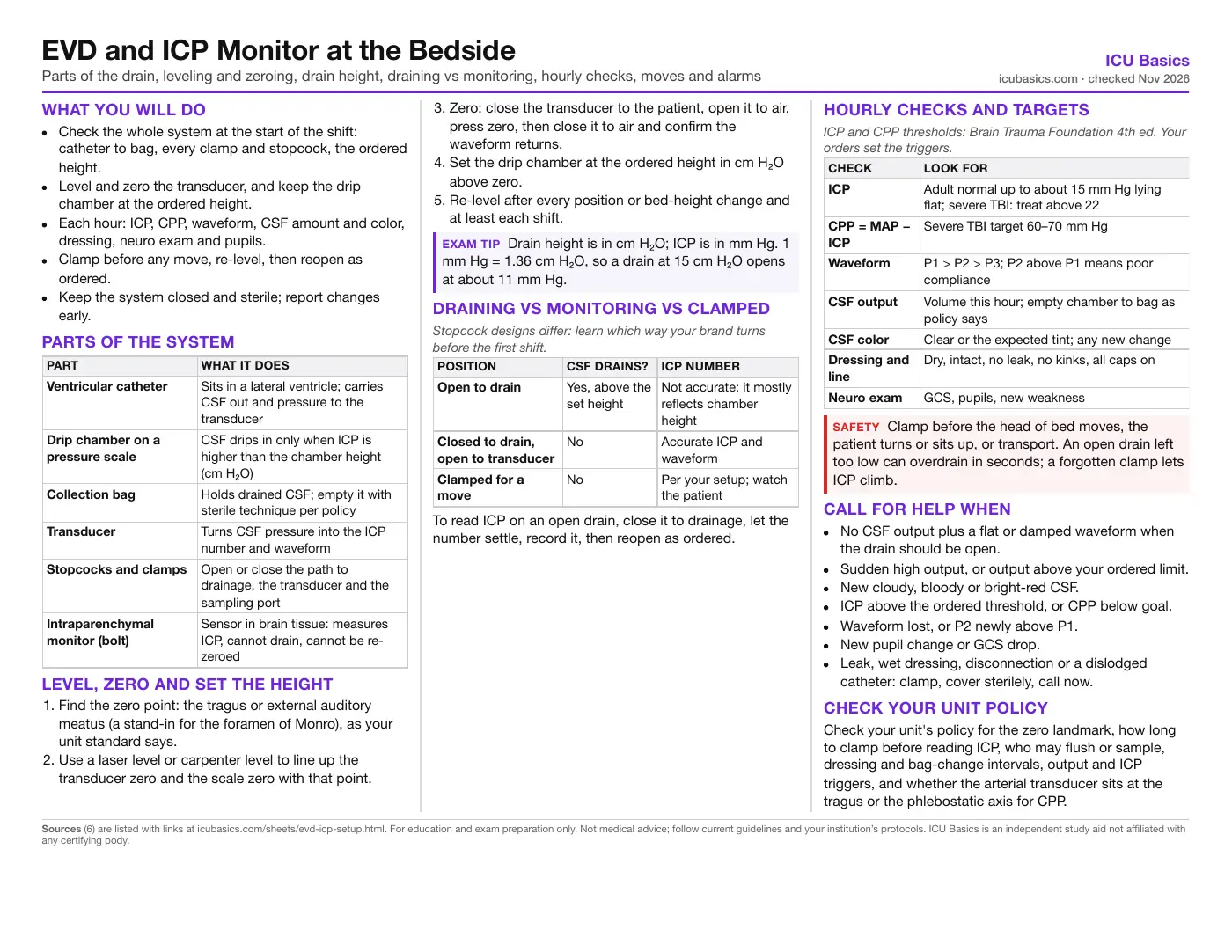
What you will do
- Check the whole system at the start of the shift: catheter to bag, every clamp and stopcock, the ordered height.
- Level and zero the transducer, and keep the drip chamber at the ordered height.
- Each hour: ICP, CPP, waveform, CSF amount and color, dressing, neuro exam and pupils.
- Clamp before any move, re-level, then reopen as ordered.
- Keep the system closed and sterile; report changes early.
Parts of the system
| Part | What it does |
|---|---|
| Ventricular catheter | Sits in a lateral ventricle; carries CSF out and pressure to the transducer |
| Drip chamber on a pressure scale | CSF drips in only when ICP is higher than the chamber height (cm H₂O) |
| Collection bag | Holds drained CSF; empty it with sterile technique per policy |
| Transducer | Turns CSF pressure into the ICP number and waveform |
| Stopcocks and clamps | Open or close the path to drainage, the transducer and the sampling port |
| Intraparenchymal monitor (bolt) | Sensor in brain tissue: measures ICP, cannot drain, cannot be re-zeroed |
Level, zero and set the height
- Find the zero point: the tragus or external auditory meatus (a stand-in for the foramen of Monro), as your unit standard says.
- Use a laser level or carpenter level to line up the transducer zero and the scale zero with that point.
- Zero: close the transducer to the patient, open it to air, press zero, then close it to air and confirm the waveform returns.
- Set the drip chamber at the ordered height in cm H₂O above zero.
- Re-level after every position or bed-height change and at least each shift.
Draining vs monitoring vs clamped
| Position | CSF drains? | ICP number |
|---|---|---|
| Open to drain | Yes, above the set height | Not accurate: it mostly reflects chamber height |
| Closed to drain, open to transducer | No | Accurate ICP and waveform |
| Clamped for a move | No | Per your setup; watch the patient |
To read ICP on an open drain, close it to drainage, let the number settle, record it, then reopen as ordered.
Hourly checks and targets
| Check | Look for |
|---|---|
| ICP | Adult normal up to about 15 mm Hg lying flat; severe TBI: treat above 22 |
| CPP = MAP − ICP | Severe TBI target 60–70 mm Hg |
| Waveform | P1 > P2 > P3; P2 above P1 means poor compliance |
| CSF output | Volume this hour; empty chamber to bag as policy says |
| CSF color | Clear or the expected tint; any new change |
| Dressing and line | Dry, intact, no leak, no kinks, all caps on |
| Neuro exam | GCS, pupils, new weakness |
Call for help when
- No CSF output plus a flat or damped waveform when the drain should be open.
- Sudden high output, or output above your ordered limit.
- New cloudy, bloody or bright-red CSF.
- ICP above the ordered threshold, or CPP below goal.
- Waveform lost, or P2 newly above P1.
- New pupil change or GCS drop.
- Leak, wet dressing, disconnection or a dislodged catheter: clamp, cover sterilely, call now.
Check your unit policy
Check your unit's policy for the zero landmark, how long to clamp before reading ICP, who may flush or sample, dressing and bag-change intervals, output and ICP triggers, and whether the arterial transducer sits at the tragus or the phlebostatic axis for CPP.
In depth
The one-page summary above is what the PDF prints. These sections go further.
How the drain decides how much CSF leaves
An EVD drains by gravity against a set height. The zero of the scale sits at the head landmark; the drip chamber is raised the ordered number of cm H₂O above it. CSF flows only while ICP pushes harder than that column. Lower the chamber and more drains; raise the patient's head without moving the chamber and the effective height falls, so CSF pours out. That is why a head-of-bed change without re-leveling causes overdrainage.
| Ordered height | Opens at about |
|---|---|
| 10 cm H₂O | 7 mm Hg |
| 15 cm H₂O | 11 mm Hg |
| 20 cm H₂O | 15 mm Hg |
Orders come in two common styles. Continuous drainage keeps the drain open at a set height, and you close it at set times to read ICP. Intermittent drainage keeps it closed with continuous ICP monitoring and opens it when ICP passes a set number. The adult brain makes CSF at roughly 20 mL an hour, so steady output well above that is a warning sign; many protocols set a report threshold around 20 mL/h, but the number is order-specific.
Reading ICP accurately
- An open drain shows a number, but it mostly reflects the chamber height. The AANN 2023 review recommends the clamped method for an accurate value and waveform.
- After closing to drainage, give the reading time to settle. Studies clamped for 5 to 15 minutes; many units read after a shorter, set time. Follow your policy and record the value the same way each time.
- Re-zero the EVD transducer when values look wrong, after disconnection and as the manufacturer or policy says. AANN found no research on zeroing technique and advises following the manufacturer.
- A damped trace with no CSF: check the patient first, then clamps and stopcocks, kinks, the ordered height and a wet air filter on the chamber.
The ICP waveform in brief
| Peak | Name | Source |
|---|---|---|
| P1 | Percussion | Arterial pulse transmitted through the choroid plexus |
| P2 | Tidal | Rebound of brain tissue; rises as compliance falls |
| P3 | Dicrotic | Aortic valve closure |
Normally P1 > P2 > P3. When P2 rises to or above P1 the brain has little room left: a small added volume will raise ICP sharply, even if the mean ICP is still in range. Report it and look for causes such as pain, agitation, fever, a kinked neck or hypercarbia. A flat trace with no peaks usually means a technical problem: a closed stopcock, clot, air or a disconnected cable.
Moving and transporting the patient
- Confirm the order allows clamping, and for how long. Some patients do not tolerate it; ask before a long trip.
- Clamp the drain (and close the chamber vent if your system calls for it) before turning, sitting up or transfer.
- Keep the drain upright; a chamber laid flat can wet the air filter and stop drainage.
- Watch the patient while clamped: neuro status, ICP if it still reads, headache or vomiting.
- When settled, re-level to the landmark, check the height, then reopen as ordered and confirm drip and waveform.
- Many units use a two-person check or checklist for the clamp-and-reopen step.
Infection prevention
- Keep the system closed. Every entry (sampling, bag change) is an infection risk; enter only with sterile technique and only as often as needed.
- Sample CSF from the proximal sampling port, never from the bag or chamber, and only when clinically indicated or ordered. Clean the port first; aspirate slowly.
- Never flush toward the patient unless your institution names you as the person who does it and there is an order; in most units only neurosurgery flushes.
- Dressing: sterile, dry and occlusive; reported change intervals vary widely, so follow policy and change it at once if wet, soiled or loose.
- Bundled care (sterile insertion, hand hygiene, minimal access, dressing standard, staff education) lowered EVD infection rates in pooled studies, about 16% before protocols vs about 5% after, from low-quality data.
- The 2016 Neurocritical Care Society consensus supports antimicrobial-impregnated catheters, one periprocedural antibiotic dose rather than continuous prophylaxis, and no routine catheter exchange.
Ventriculitis can hide behind fever and blood in the CSF that are already there. Watch the trend: CSF turning cloudy, new fever, a worsening exam. Report any change in CSF color or clarity; blood-tinged CSF after a bleed is expected and should slowly clear.
Intraparenchymal monitors
- A fiberoptic or strain-gauge sensor placed into brain tissue, often through a bolt in the skull.
- Measures ICP continuously; cannot drain CSF.
- Zeroed once, before insertion. After insertion it cannot be re-zeroed, and drift grows the longer it stays in.
- The sensor reads at its tip, so there is no external transducer to level.
- Infection risk is lower than with an EVD. Protect the cable and connector; do not kink or pull it.
- A reading that does not fit the patient: check the connection and the patient, then call; the team may compare it with imaging or replace the device.
Targets and evidence
The Brain Trauma Foundation 4th edition (2016, still current) recommends treating ICP above 22 mm Hg in severe TBI and a CPP target of 60 to 70 mm Hg. Those numbers come from TBI; many non-TBI protocols treat ICP above 20. CPP depends on where the arterial transducer is leveled: AANN notes practice varies between the tragus and the phlebostatic axis, which changes MAP and so CPP. Use your unit's standard every time.
There is no proven best external zero point. Tragus, external auditory meatus and other spots are all used; consistency on a unit matters more than which one.
Related cheat sheets
- Your First Weeks in the ICUHow ICU orientation works, getting the most from your preceptor, organizing a shift, and speaking up for safety
- ICU Report Sheet: SBAR Brain Sheet for NursesA typical ICU shift hour by hour, a printable system-by-system report layout, SBAR handoff and what to check together at the bedside
- Drawing ICU Labs: Order of Draw, Cultures and Timed LabsOrder of draw, line draws without contamination, blood cultures, blood gases, labeling and the labs that run on a clock
- Daily Bedside Screens: Skin, Falls, Swallow and WithdrawalPressure injury risk, fall risk, the post-extubation swallow screen and CIWA-Ar, and what to do with each result
- Early Mobility in the ICUWhy it matters, a step-by-step progression, the safety screen before and stop criteria during a session, team roles, equipment and documentation
- Leaving the Unit: CT, MRI, IR, Cath Lab and ORIs the trip worth it, who goes, what travels, monitoring and handoff, then what to check at each destination
Calculators for this topic
Sources
- Tran DS, Supan E, Young A, Ricke D, Censullo J. Evidence-Based Clinical Review: Intracranial Monitoring. American Association of Neuroscience Nurses; 2023 (originated 2011 as an AANN Clinical Practice Guideline; revised 2012 and 2014). Link
- American Association of Neuroscience Nurses. A Nurse's Guide to Maintaining External Ventricular Drains. Link
- Fried HI, Nathan BR, Rowe AS, et al. The Insertion and Management of External Ventricular Drains: An Evidence-Based Consensus Statement. A Statement for Healthcare Professionals from the Neurocritical Care Society. Neurocrit Care. 2016;24(1):61–81. Link
- Carney N, Totten AM, O'Reilly C, et al. Guidelines for the Management of Severe Traumatic Brain Injury, Fourth Edition. Brain Trauma Foundation. Neurosurgery. 2017;80(1):6–15. Link
- Vieira TW, Sakamoto VTM, Araujo BR, et al. External Ventricular Drains: Development and Evaluation of a Nursing Clinical Practice Guideline. Nurs Rep. 2022. Link
- Farkas J. External ventricular drain (EVD). Internet Book of Critical Care (EMCrit). Link