 ICU Basics
Cheat Sheet
Drawing ICU Labs: Order of Draw, Cultures and Timed Labs
Order of draw, line draws without contamination, blood cultures, blood gases, labeling and the labs that run on a clock
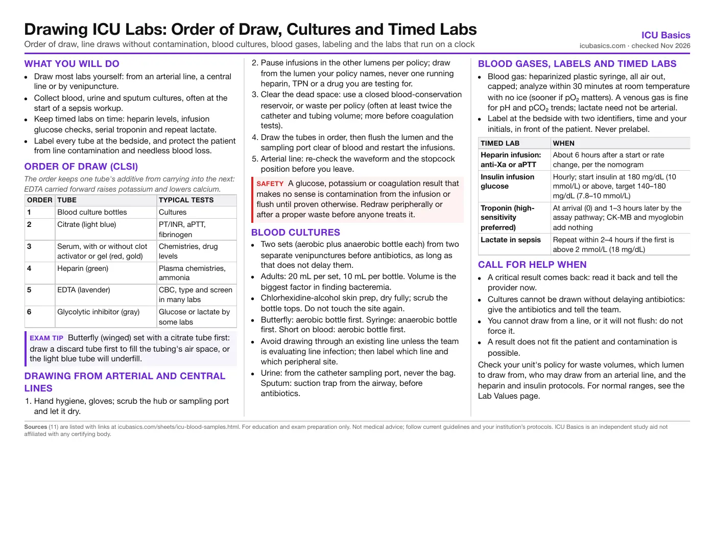
What you will do
- Draw most labs yourself: from an arterial line, a central line or by venipuncture.
- Collect blood, urine and sputum cultures, often at the start of a sepsis workup.
- Keep timed labs on time: heparin levels, infusion glucose checks, serial troponin and repeat lactate.
- Label every tube at the bedside, and protect the patient from line contamination and needless blood loss.
Order of draw (CLSI)
| Order | Tube | Typical tests |
|---|---|---|
| 1 | Blood culture bottles | Cultures |
| 2 | Citrate (light blue) | PT/INR, aPTT, fibrinogen |
| 3 | Serum, with or without clot activator or gel (red, gold) | Chemistries, drug levels |
| 4 | Heparin (green) | Plasma chemistries, ammonia |
| 5 | EDTA (lavender) | CBC, type and screen in many labs |
| 6 | Glycolytic inhibitor (gray) | Glucose or lactate by some labs |
Drawing from arterial and central lines
- Hand hygiene, gloves; scrub the hub or sampling port and let it dry.
- Pause infusions in the other lumens per policy; draw from the lumen your policy names, never one running heparin, TPN or a drug you are testing for.
- Clear the dead space: use a closed blood-conservation reservoir, or waste per policy (often at least twice the catheter and tubing volume; more before coagulation tests).
- Draw the tubes in order, then flush the lumen and the sampling port clear of blood and restart the infusions.
- Arterial line: re-check the waveform and the stopcock position before you leave.
Blood cultures
- Two sets (aerobic plus anaerobic bottle each) from two separate venipunctures before antibiotics, as long as that does not delay them.
- Adults: 20 mL per set, 10 mL per bottle. Volume is the biggest factor in finding bacteremia.
- Chlorhexidine-alcohol skin prep, dry fully; scrub the bottle tops. Do not touch the site again.
- Butterfly: aerobic bottle first. Syringe: anaerobic bottle first. Short on blood: aerobic bottle first.
- Avoid drawing through an existing line unless the team is evaluating line infection; then label which line and which peripheral site.
- Urine: from the catheter sampling port, never the bag. Sputum: suction trap from the airway, before antibiotics.
Blood gases, labels and timed labs
- Blood gas: heparinized plastic syringe, all air out, capped; analyze within 30 minutes at room temperature with no ice (sooner if pO₂ matters). A venous gas is fine for pH and pCO₂ trends; lactate need not be arterial.
- Label at the bedside with two identifiers, time and your initials, in front of the patient. Never prelabel.
| Timed lab | When |
|---|---|
| Heparin infusion: anti-Xa or aPTT | About 6 hours after a start or rate change, per the nomogram |
| Insulin infusion glucose | Hourly; start insulin at 180 mg/dL (10 mmol/L) or above, target 140–180 mg/dL (7.8–10 mmol/L) |
| Troponin (high-sensitivity preferred) | At arrival (0) and 1–3 hours later by the assay pathway; CK-MB and myoglobin add nothing |
| Lactate in sepsis | Repeat within 2–4 hours if the first is above 2 mmol/L (18 mg/dL) |
Call for help when
- A critical result comes back: read it back and tell the provider now.
- Cultures cannot be drawn without delaying antibiotics: give the antibiotics and tell the team.
- You cannot draw from a line, or it will not flush: do not force it.
- A result does not fit the patient and contamination is possible.
Check your unit's policy for waste volumes, which lumen to draw from, who may draw from an arterial line, and the heparin and insulin protocols. For normal ranges, see the Lab Values page.
In depth
The one-page summary above is what the PDF prints. These sections go further.
Why the order of draw matters
Every tube additive can contaminate the next tube through the needle or holder. Cultures go first so the sterile bottles meet the needle before anything else. Citrate goes next because a clot activator or heparin carried into it changes coagulation times. EDTA comes late because it binds calcium and carries potassium (it is a potassium salt): an EDTA carryover can show a falsely high potassium and falsely low calcium. Fluoride/oxalate (gray) comes last because it damages cells and raises potassium and sodium in later tubes.
Studies with closed, modern systems show little carryover when technique is good, but CLSI keeps the order because it costs nothing and a single error can trigger a dangerous treatment, such as treating a false hyperkalemia.
- Fill each tube to its line. A short citrate tube has too much anticoagulant for the blood and prolongs the PT and aPTT.
- Invert each tube gently the number of times the maker states; do not shake.
- Very high hematocrit (above 55%) needs a citrate volume adjustment; the lab will tell you.
Line draws: contamination and blood loss
The commonest ICU lab errors come from line draws. Infusions in a neighboring lumen can reach the sampling lumen, and the flush solution sits in the catheter until it is cleared. Dextrose raises glucose, potassium or saline infusions change electrolytes, and heparin flush prolongs the aPTT and can make anti-Xa levels useless.
| Result pattern | Suspect |
|---|---|
| Very high glucose with normal other values | Dextrose or TPN contamination |
| High potassium with no ECG change | Potassium infusion contamination or a hemolyzed sample |
| Wildly prolonged aPTT or high anti-Xa | Heparin from the line or flush |
| Low hemoglobin and electrolytes all at once | Dilution from inadequate waste |
- Closed blood-conservation systems on arterial lines return the waste volume to the patient and cut iatrogenic blood loss.
- Small-volume (pediatric) tubes, batching labs and stopping standing daily orders that no longer change care all reduce ICU anemia and transfusion.
- Waste volume: reviews support about twice the dead space for most tests and up to about six times before coagulation tests from a heparinized line. Follow your policy.
- Never draw from a peripheral IV above an infusion, or from the same arm as an infusion if you can avoid it; if you must, stop the infusion and draw below it per policy.
Blood cultures in detail
Blood culture yield depends mostly on volume. For adults, the IDSA/ASM 2024 guide and CLSI M47 call for 20 to 30 mL per set and two to four sets for a bacteremia workup, usually two sets of 10 mL per bottle. Underfilled bottles miss organisms; overfilled bottles can give false positives on some systems.
- Two sets from separate venipunctures let the lab tell a contaminant (coagulase-negative staph in one set) from true bacteremia (the same organism in both).
- Contamination is driven by skin prep: use chlorhexidine-alcohol or the agent your lab chooses, let it dry, and do not palpate the site after prep unless wearing sterile gloves.
- Blood drawn through an existing catheter is more often contaminated. Draw through a line only when evaluating catheter-related bloodstream infection: then one set through the catheter and one peripheral set, labeled with the source and drawn close together in time.
- Get the bottles to the lab promptly; do not refrigerate.
- Sepsis: Surviving Sepsis 2026 strongly recommends blood cultures as soon as possible, ideally before antibiotics. SSC 2021 set the limit: cultures first only if they add no substantial delay (under about 45 minutes). In septic shock or probable sepsis, antibiotics ideally within 1 hour come first.
- If you get less than a full set, put the blood in the aerobic bottle first; most bloodstream organisms grow there.
Urine, sputum and other cultures
- Catheter urine: clamp the tubing below the port briefly if needed, scrub the sampling port, aspirate with a sterile syringe. Never from the drainage bag. A catheter in place for a long time may be changed first per policy so the sample reflects the bladder, not the biofilm.
- Send urine for culture only when there are signs of infection; asymptomatic bacteriuria in a catheterized patient is not usually treated.
- Sputum in an intubated patient: endotracheal aspirate in an in-line sterile trap; label as tracheal aspirate. The provider may do a bronchoscopic or mini-BAL sample.
- Send all culture specimens promptly; refrigerate urine if transport will be delayed, per your lab.
Blood gases: arterial or venous
Use a preheparinized plastic syringe, expel air bubbles at once (air raises pO₂ and lowers pCO₂), cap and mix. CLSI C46: plastic syringes stay at room temperature and are analyzed within 30 minutes; samples for special studies (such as a shunt study) within about 5 minutes. A very high white cell or platelet count consumes oxygen in the syringe, so run those at once. Ice was for glass syringes; cold plastic lets gas move through the wall.
- Venous pH runs a little lower and pCO₂ a little higher than arterial; a venous gas works for trending pH and pCO₂ but not oxygenation.
- Lactate from venous blood is accepted; an arterial stick only for lactate is not needed.
- Wait per protocol after a ventilator change before drawing the gas, so the result reflects the new settings.
- Note FiO₂, ventilator settings and temperature on the request.
Timed labs: why the clock matters
- Heparin: unfractionated heparin reaches a new steady state about 6 hours after a rate change, so levels drawn early mislead the nomogram. Draw from the arm without the heparin, or from a line not used for heparin. Anti-Xa is less affected by factor levels and acute-phase proteins than the aPTT.
- Insulin infusion: SCCM 2024, ADA 2026 and Surviving Sepsis 2026 start insulin for persistent glucose 180 mg/dL (10 mmol/L) or above; SCCM and ADA aim for 140–180 mg/dL; tighter targets raised hypoglycemia and deaths (NICE-SUGAR). Check hourly while the rate is changing; some protocols space checks to every 2 hours once stable. Fingerstick values can be unreliable in shock, edema or on vasopressors; use arterial or venous blood on a meter approved for it, per policy.
- Troponin: AHA/ACC 2021 prefers high-sensitivity troponin with a clinical decision pathway: 0 and 1 hour, or 0 and 2 to 3 hours, depending on the assay. ESC 2023 prefers 0 and 1 hour, with 0 and 2 hours as the alternative. With a conventional assay, repeat at 3 to 6 hours. CK-MB and myoglobin are not useful for diagnosing MI. Record the exact draw time; the pathway uses the change between values.
- Lactate: the Surviving Sepsis Hour-1 bundle measures lactate and remeasures within 2 to 4 hours if the first is above 2 mmol/L (18 mg/dL). Surviving Sepsis 2026 suggests measuring lactate and using serial values to guide resuscitation, but not giving fluid just to normalize lactate.
Labeling and specimen safety
- Two identifiers (name and date of birth, or medical record number) checked against the armband at the bedside.
- Label in the patient's presence immediately after the draw; never take unlabeled tubes out of the room or label ahead of time.
- Wrong-blood-in-tube errors on a type and screen can cause a fatal transfusion reaction; many hospitals need a second sample or a separate draw for a first type.
- Use scanning tools if your hospital has them; they reduce but do not remove identification errors.
Related cheat sheets
- Your First Weeks in the ICUHow ICU orientation works, getting the most from your preceptor, organizing a shift, and speaking up for safety
- ICU Report Sheet: SBAR Brain Sheet for NursesA typical ICU shift hour by hour, a printable system-by-system report layout, SBAR handoff and what to check together at the bedside
- Daily Bedside Screens: Skin, Falls, Swallow and WithdrawalPressure injury risk, fall risk, the post-extubation swallow screen and CIWA-Ar, and what to do with each result
- Early Mobility in the ICUWhy it matters, a step-by-step progression, the safety screen before and stop criteria during a session, team roles, equipment and documentation
- Leaving the Unit: CT, MRI, IR, Cath Lab and ORIs the trip worth it, who goes, what travels, monitoring and handoff, then what to check at each destination
- Central Lines: Care, CLABSI Prevention and TroubleshootingLine types, tip check, the maintenance bundle, draws and flushes, occlusion, air embolism, infection and removal
Calculators for this topic
Sources
- Clinical and Laboratory Standards Institute. PRE02: Collection of Diagnostic Venous Blood Specimens. 8th ed. (replaces GP41-Ed7, 2017). Wayne, PA: CLSI; 2025. Link
- Clinical and Laboratory Standards Institute. M47: Principles and Procedures for Blood Cultures. 2nd ed. Wayne, PA: CLSI; 2022. Link
- Clinical and Laboratory Standards Institute. C46: Blood Gas and pH Analysis and Related Measurements. 2nd ed. Wayne, PA: CLSI; 2009. Link
- Miller JM, Binnicker MJ, Campbell S, et al. Guide to Utilization of the Microbiology Laboratory for Diagnosis of Infectious Diseases: 2024 Update by IDSA and ASM. Clin Infect Dis. 2024; ciae104. Link
- Evans L, Rhodes A, Alhazzani W, et al. Surviving Sepsis Campaign: International Guidelines for Management of Sepsis and Septic Shock 2021. Intensive Care Med. 2021;47:1181–1247. Link
- Prescott HC, Antonelli M, Alhazzani W, et al. Surviving Sepsis Campaign: International Guidelines for Management of Sepsis and Septic Shock 2026. Crit Care Med. 2026;54(4):725–812. doi:10.1097/CCM.0000000000007075. Link
- Levy MM, Evans LE, Rhodes A. The Surviving Sepsis Campaign Bundle: 2018 Update. Crit Care Med. 2018;46:997–1000. Link
- Gulati M, Levy PD, Mukherjee D, et al. 2021 AHA/ACC/ASE/CHEST/SAEM/SCCT/SCMR Guideline for the Evaluation and Diagnosis of Chest Pain. Circulation. 2021;144:e368–e454. Link
- Byrne RA, Rossello X, Coughlan JJ, et al. 2023 ESC Guidelines for the management of acute coronary syndromes. Eur Heart J. 2023;44:3720–3826. Link
- American Diabetes Association Professional Practice Committee. 16. Diabetes Care in the Hospital: Standards of Care in Diabetes—2026. Diabetes Care. 2026;49(Suppl 1):S339–S355. Link
- Honarmand K, Sirimaturos M, Hirshberg EL, et al. Society of Critical Care Medicine Guidelines on Glycemic Control for Critically Ill Children and Adults 2024. Crit Care Med. 2024;52:e161–e181. Link