 ICU Basics
Cheat Sheet
Early Mobility in the ICU
Why it matters, a step-by-step progression, the safety screen before and stop criteria during a session, team roles, equipment and documentation
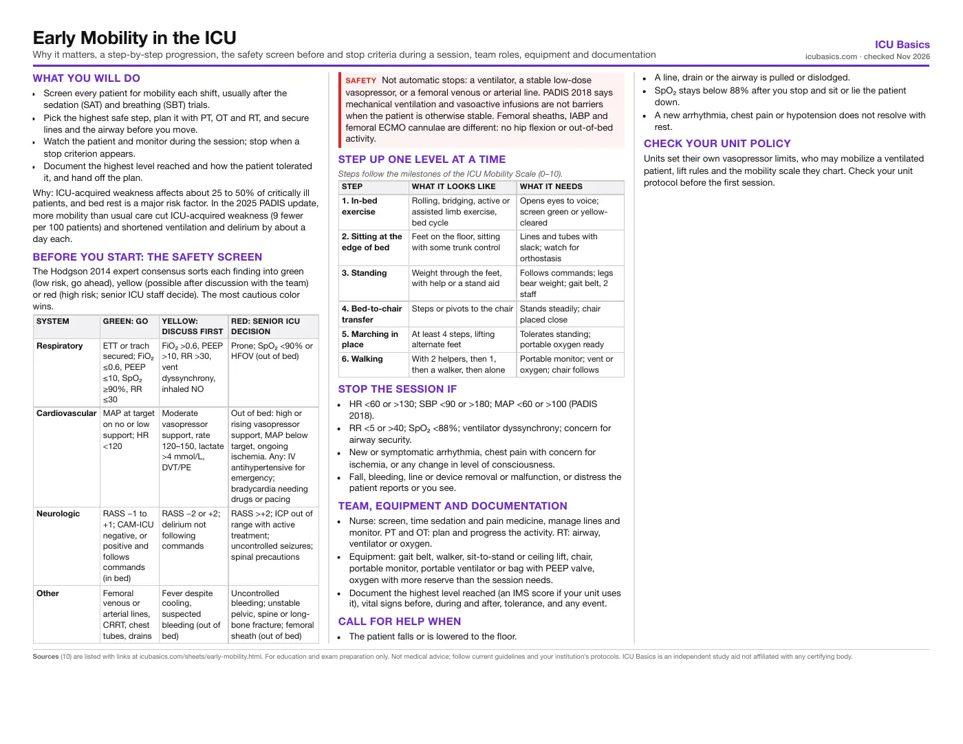
What you will do
- Screen every patient for mobility each shift, usually after the sedation (SAT) and breathing (SBT) trials.
- Pick the highest safe step, plan it with PT, OT and RT, and secure lines and the airway before you move.
- Watch the patient and monitor during the session; stop when a stop criterion appears.
- Document the highest level reached and how the patient tolerated it, and hand off the plan.
Why: ICU-acquired weakness affects about 25 to 50% of critically ill patients, and bed rest is a major risk factor. In the 2025 PADIS update, more mobility than usual care cut ICU-acquired weakness (9 fewer per 100 patients) and shortened ventilation and delirium by about a day each.
Before you start: the safety screen
The Hodgson 2014 expert consensus sorts each finding into green (low risk, go ahead), yellow (possible after discussion with the team) or red (high risk; senior ICU staff decide). The most cautious color wins.
| System | Green: go | Yellow: discuss first | Red: senior ICU decision |
|---|---|---|---|
| Respiratory | ETT or trach secured; FiO₂ ≤0.6, PEEP ≤10, SpO₂ ≥90%, RR ≤30 | FiO₂ >0.6, PEEP >10, RR >30, vent dyssynchrony, inhaled NO | Prone; SpO₂ <90% or HFOV (out of bed) |
| Cardiovascular | MAP at target on no or low support; HR <120 | Moderate vasopressor support, rate 120–150, lactate >4 mmol/L, DVT/PE | Out of bed: high or rising vasopressor support, MAP below target, ongoing ischemia. Any: IV antihypertensive for emergency; bradycardia needing drugs or pacing |
| Neurologic | RASS −1 to +1; CAM-ICU negative, or positive and follows commands (in bed) | RASS −2 or +2; delirium not following commands | RASS >+2; ICP out of range with active treatment; uncontrolled seizures; spinal precautions |
| Other | Femoral venous or arterial lines, CRRT, chest tubes, drains | Fever despite cooling, suspected bleeding (out of bed) | Uncontrolled bleeding; unstable pelvic, spine or long-bone fracture; femoral sheath (out of bed) |
Step up one level at a time
| Step | What it looks like | What it needs |
|---|---|---|
| 1. In-bed exercise | Rolling, bridging, active or assisted limb exercise, bed cycle | Opens eyes to voice; screen green or yellow-cleared |
| 2. Sitting at the edge of bed | Feet on the floor, sitting with some trunk control | Lines and tubes with slack; watch for orthostasis |
| 3. Standing | Weight through the feet, with help or a stand aid | Follows commands; legs bear weight; gait belt, 2 staff |
| 4. Bed-to-chair transfer | Steps or pivots to the chair | Stands steadily; chair placed close |
| 5. Marching in place | At least 4 steps, lifting alternate feet | Tolerates standing; portable oxygen ready |
| 6. Walking | With 2 helpers, then 1, then a walker, then alone | Portable monitor; vent or oxygen; chair follows |
Stop the session if
- HR <60 or >130; SBP <90 or >180; MAP <60 or >100 (PADIS 2018).
- RR <5 or >40; SpO₂ <88%; ventilator dyssynchrony; concern for airway security.
- New or symptomatic arrhythmia, chest pain with concern for ischemia, or any change in level of consciousness.
- Fall, bleeding, line or device removal or malfunction, or distress the patient reports or you see.
Team, equipment and documentation
- Nurse: screen, time sedation and pain medicine, manage lines and monitor. PT and OT: plan and progress the activity. RT: airway, ventilator or oxygen.
- Equipment: gait belt, walker, sit-to-stand or ceiling lift, chair, portable monitor, portable ventilator or bag with PEEP valve, oxygen with more reserve than the session needs.
- Document the highest level reached (an IMS score if your unit uses it), vital signs before, during and after, tolerance, and any event.
Call for help when
- The patient falls or is lowered to the floor.
- A line, drain or the airway is pulled or dislodged.
- SpO₂ stays below 88% after you stop and sit or lie the patient down.
- A new arrhythmia, chest pain or hypotension does not resolve with rest.
Check your unit policy
Units set their own vasopressor limits, who may mobilize a ventilated patient, lift rules and the mobility scale they chart. Check your unit protocol before the first session.
In depth
The one-page summary above is what the PDF prints. These sections go further.
The evidence: start early, match the dose to the patient
PADIS 2018 suggested rehabilitation or mobilization for critically ill adults (conditional, low-quality evidence). Pooled trials showed more strength at ICU discharge (MRC sum score +6.2 points) and about 1.3 fewer ventilator days, with no change in hospital mortality. Serious safety events were rare: 15 across more than 12,200 sessions, most often respiratory (desaturation needing more oxygen, unplanned extubation).
The 2025 focused update (58 trials, 8,038 patients) suggests enhanced mobilization over usual care (conditional, moderate certainty). "Enhanced" meant anything more than a unit already did: earlier, twice daily, protocolized, longer, cycling or stepping. It reduced ICU-acquired weakness (RR 0.77), and may shorten delirium (about 1.3 days), ventilation (1.1 days) and ICU stay (1 day). Arrhythmias may be slightly more common (about 1 more per 100); line removal, unplanned extubation and hypotension were not clearly increased. The panel could not set an ideal dose, timing or method.
Schweickert 2009 paired PT and OT with daily sedation interruption early in ventilation: 59% vs 35% returned to independent function by hospital discharge, with fewer delirium days. The TEAM trial (Hodgson 2022, 750 ventilated adults) pushed harder: 20.8 vs 8.8 minutes of active mobilization a day. Days alive and out of hospital at 180 days were no different (143 vs 145), and adverse events possibly from mobilization (arrhythmia, blood pressure change, desaturation) were more common, 9.2% vs 4.1%.
The full traffic-light screen (Hodgson 2014)
The consensus covers active mobilization of mechanically ventilated adults, split into in-bed and out-of-bed exercise. It did not address passive range of motion or monitoring during a session. Before each session, someone named by the unit confirms the airway is positioned and secured and that oxygen on hand outlasts the planned activity.
| Finding | In bed | Out of bed |
|---|---|---|
| ETT or tracheostomy | Green | Green |
| FiO₂ >0.6; PEEP >10; RR >30; dyssynchrony | Yellow | Yellow |
| SpO₂ <90% | Yellow | Red |
| HFOV | Yellow | Red |
| Inhaled nitric oxide or prostacyclin | Yellow | Yellow |
| Prone positioning | Red | Red |
| MAP at target, no or low support | Green | Green |
| MAP at target, moderate support | Yellow | Yellow |
| MAP at target, high support | Yellow | Red |
| MAP below target despite support, or with symptoms | Yellow | Red |
| IV antihypertensive for hypertensive emergency | Red | Red |
| Bradycardia needing drugs or emergency pacing | Red | Red |
| Pacemaker-dependent rhythm | Yellow | Red |
| Stable tachyarrhythmia, rate >150 / 120–150 / <120 | Yellow / Yellow / Green | Red / Yellow / Green |
| Femoral IABP; femoral or subclavian ECMO | Green | Red |
| Single bicaval dual-lumen ECMO cannula | Green | Yellow |
| VAD | Green | Green |
| PA catheter or continuous CO monitor | Green | Yellow |
| Lactate >4 mmol/L; known DVT or PE | Yellow | Yellow |
| Ongoing cardiac ischemia | Yellow | Red |
| RASS −1 to +1 | Green | Green |
| RASS −2 or +2 | Yellow | Yellow |
| RASS below −2 | Yellow | Red |
| RASS above +2 | Red | Red |
| CAM-ICU positive, follows simple commands | Green | Yellow |
| CAM-ICU positive, not following commands | Yellow | Yellow |
| ICP out of range with active treatment | Red | Red |
| ICP monitor, no active treatment; craniectomy | Green | Yellow |
| Open (unclamped) lumbar drain | Green | Red |
| Spinal precautions before clearance; uncontrolled seizures | Red | Red |
| Unclipped aneurysm after SAH; vasospasm after clipping | Green | Yellow |
| Unstable pelvic, spinal or long-bone fracture | Yellow | Red |
| Large open chest or abdominal wound | Green | Red |
| Uncontrolled active bleeding | Red | Red |
| Suspected bleeding or high bleeding risk | Green | Yellow |
| Fever despite cooling; active hypothermia management | Yellow | Yellow |
| Femoral venous or arterial catheter; CRRT incl. femoral dialysis catheter | Green | Green |
| Femoral sheath | Yellow | Red |
| NG tube, CVC, chest tube, wound drain, urinary catheter | Green | Green |
Vasopressors, femoral lines and devices
Hodgson 2014 could not agree on a vasopressor dose below which mobility is safe. The panel did agree that the drug itself is not an absolute contraindication; what matters is the absolute dose, whether the dose is rising (a reason for caution or to hold), and whether the patient is well perfused. Each ICU sets its own threshold. In practice: a patient at MAP goal on a stable low dose with warm skin, normal mentation and a falling lactate can usually do in-bed and often out-of-bed work; a patient whose norepinephrine was just increased cannot.
Femoral venous and arterial catheters, and CRRT through a femoral dialysis catheter, are green for in- and out-of-bed activity in both Hodgson 2014 and PADIS 2018. Secure the line, keep slack in the tubing, watch the site and pressure waveform afterward, and coordinate with dialysis so a circuit pause is planned rather than an alarm. Femoral sheaths, femoral IABPs and femoral ECMO cannulae are different: hip flexion can kink or dislodge them, so in-bed exercise may need changing and out-of-bed activity is off the table.
Running a session: before, during, after
- Before: run the screen, check the plan with the provider and therapists, and time analgesia so pain peaks are covered. Ask RT to bring a portable ventilator or a bag with PEEP valve and confirm the oxygen supply.
- Organize the lines: move pumps and the monitor to the side you will go, add extension tubing as needed, clamp or drain per policy (for example a urinary catheter bag kept below the bladder), and assign one person to the airway.
- Brief the patient: what you will do, how they can help, and a signal to stop.
- Dangle first: raise the head of bed, then sit at the edge of bed and recheck vital signs and symptoms before standing.
- During: watch the face and the monitor, not only the numbers. Coach breathing, and stop at the first stop criterion. Keep the chair or bed close.
- After: return to a safe position, recheck vital signs, waveforms and line sites, reconnect the ventilator settings or oxygen, and document.
The ICU Mobility Scale
The ICU Mobility Scale (IMS, Hodgson 2014) records the highest level a patient reached, from 0 to 10. Most clinicians in the original study scored it in under a minute, and agreement between nurses and physiotherapists was good (weighted kappa about 0.7). Higher IMS at ICU discharge goes with better 90-day survival and discharge home, and a change of about 3 points is clinically meaningful (Tipping 2018).
| IMS | Highest level reached |
|---|---|
| 0 | Nothing: lying in bed, passively rolled or exercised |
| 1 | Active exercise or activity in bed, not over the edge |
| 2 | Passively moved to a chair by lift or slide, no standing |
| 3 | Sitting over the edge of the bed with some trunk control |
| 4 | Standing, with or without help or a stand aid |
| 5 | Transferring bed to chair by stepping or shuffling |
| 6 | Marching on the spot, at least 4 steps |
| 7 | Walking at least 16 ft (5 m) with 2 or more helpers |
| 8 | Walking with 1 helper |
| 9 | Walking independently with a gait aid |
| 10 | Walking independently without a gait aid |
Other tools units use include the AACN Early Progressive Mobility Protocol, the Johns Hopkins Highest Level of Mobility scale and bedside mobility assessment tools. Use whichever your unit charts, and chart the same way every time so the trend is real.
Common barriers and what to try
| Barrier | What to try |
|---|---|
| Too sedated (RASS below −2) | Pair the session with the SAT; analgesia first, lightest sedation that works |
| Delirium | Calm, short commands; family at the bedside; delirium is not a stop if they follow simple commands |
| Pain | Premedicate and time the session to the peak effect; splint incisions |
| Orthostatic hypotension | Raise the head of bed in stages; dangle longer; compression devices per order |
| Desaturation with effort | Ask RT about a temporary FiO₂ or support increase; rest breaks; stay in bed if it persists |
| Fear or low motivation | Set a goal with the patient; explain each step; celebrate the gain |
| Short-staffed | In-bed work and edge of bed still count; schedule with PT and OT at rounds |
Nursing care and hand-off
- Report the IMS level, tolerance and the next goal at rounds and at shift change.
- Keep the patient upright in the chair as long as tolerated, with call light and suction in reach, and a plan for getting back to bed.
- Inspect skin and pressure points after sitting; reposition on schedule.
- Mobility, light sedation, delirium screening and family engagement work together; a gain in one helps the others.
- Ask about a post-ICU follow-up or rehabilitation referral for patients with lasting weakness.
Related cheat sheets
- Your First Weeks in the ICUHow ICU orientation works, getting the most from your preceptor, organizing a shift, and speaking up for safety
- ICU Report Sheet: SBAR Brain Sheet for NursesA typical ICU shift hour by hour, a printable system-by-system report layout, SBAR handoff and what to check together at the bedside
- Drawing ICU Labs: Order of Draw, Cultures and Timed LabsOrder of draw, line draws without contamination, blood cultures, blood gases, labeling and the labs that run on a clock
- Daily Bedside Screens: Skin, Falls, Swallow and WithdrawalPressure injury risk, fall risk, the post-extubation swallow screen and CIWA-Ar, and what to do with each result
- Leaving the Unit: CT, MRI, IR, Cath Lab and ORIs the trip worth it, who goes, what travels, monitoring and handoff, then what to check at each destination
- Central Lines: Care, CLABSI Prevention and TroubleshootingLine types, tip check, the maintenance bundle, draws and flushes, occlusion, air embolism, infection and removal
Calculators for this topic
Sources
- Devlin JW, Skrobik Y, Gélinas C, et al. Clinical practice guidelines for the prevention and management of pain, agitation/sedation, delirium, immobility, and sleep disruption in adult patients in the ICU (PADIS). Crit Care Med. 2018;46(9):e825–e873. Link
- Lewis K, Balas MC, Stollings JL, et al. A focused update to the clinical practice guidelines for the prevention and management of pain, anxiety, agitation/sedation, delirium, immobility, and sleep disruption in adult patients in the ICU. Crit Care Med. 2025;53(3):e711–e727. Link
- Hodgson CL, Stiller K, Needham DM, et al. Expert consensus and recommendations on safety criteria for active mobilization of mechanically ventilated critically ill adults. Crit Care. 2014;18(6):658. Link
- Hodgson C, Needham D, Haines K, et al. Feasibility and inter-rater reliability of the ICU Mobility Scale. Heart Lung. 2014;43(1):19–24. Link
- Tipping CJ, Bailey MJ, Bellomo R, et al. The ICU Mobility Scale has construct and predictive validity and is responsive. Ann Am Thorac Soc. 2016;13(6):887–893. Link
- Tipping CJ, Holland AE, Harrold M, et al. The minimal important difference of the ICU mobility scale. Heart Lung. 2018;47(5):497–501. Link
- TEAM Study Investigators and the ANZICS Clinical Trials Group; Hodgson CL, Bailey M, Bellomo R, et al. Early active mobilization during mechanical ventilation in the ICU. N Engl J Med. 2022;387(19):1747–1758. Link
- Schweickert WD, Pohlman MC, Pohlman AS, et al. Early physical and occupational therapy in mechanically ventilated, critically ill patients: a randomised controlled trial. Lancet. 2009;373(9678):1874–1882. Link
- Society of Critical Care Medicine. ICU Liberation Bundle (A-F): E element, early mobility and exercise. Link
- American Association of Critical-Care Nurses. Implementing early progressive mobility (CSI project resources and the AACN Early Progressive Mobility Protocol). Link