 ICU Basics
Cheat Sheet
Leaving the Unit: CT, MRI, IR, Cath Lab and OR
Is the trip worth it, who goes, what travels, monitoring and handoff, then what to check at each destination
What you will do
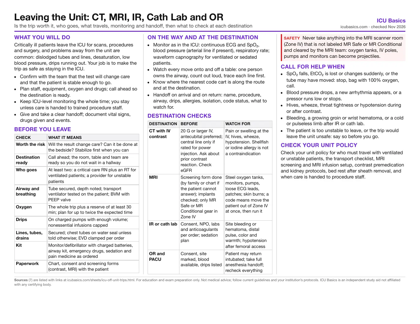
Critically ill patients leave the ICU for scans, procedures and surgery, and problems away from the unit are common: dislodged tubes and lines, desaturation, low blood pressure, drips running out. Your job is to make the trip as safe as staying in the ICU.
- Confirm with the team that the test will change care and that the patient is stable enough to go.
- Plan staff, equipment, oxygen and drugs; call ahead so the destination is ready.
- Keep ICU-level monitoring the whole time; you stay unless care is handed to trained procedure staff.
- Give and take a clear handoff; document vital signs, drugs given and events.
Before you leave
| Check | What it means |
|---|---|
| Worth the risk | Will the result change care? Can it be done at the bedside? Stabilize first when you can |
| Destination ready | Call ahead; the room, table and team are ready so you do not wait in a hallway |
| Who goes | At least two: a critical care RN plus an RT for ventilated patients; a provider for unstable patients |
| Airway and breathing | Tube secured, depth noted; transport ventilator tested on the patient; BVM with PEEP valve |
| Oxygen | The whole trip plus a reserve of at least 30 min; plan for up to twice the expected time |
| Drips | On charged pumps with enough volume; nonessential infusions capped |
| Lines, tubes, drains | Secured; chest tubes on water seal unless told otherwise; EVD clamped per order |
| Kit | Monitor/defibrillator with charged batteries, airway kit, emergency drugs, sedation and pain medicine as ordered |
| Paperwork | Chart, consent and screening forms (contrast, MRI) with the patient |
On the way and at the destination
- Monitor as in the ICU: continuous ECG and SpO₂, blood pressure (arterial line if present), respiratory rate; waveform capnography for ventilated or sedated patients.
- Watch every move onto and off a table: one person owns the airway, count out loud, trace each line first.
- Know where the nearest code cart is along the route and at the destination.
- Handoff on arrival and on return: name, procedure, airway, drips, allergies, isolation, code status, what to watch for.
Destination checks
| Destination | Before | Watch for |
|---|---|---|
| CT with IV contrast | 20 G or larger IV, antecubital preferred; central line only if rated for power injection. Ask about prior contrast reaction. Check eGFR | Pain or swelling at the IV, hives, wheeze, hypotension. Shellfish or iodine allergy is not a contraindication |
| MRI | Screening form done (by family or chart if the patient cannot answer); implants checked; only MR Safe or MR Conditional gear in Zone IV | Steel oxygen tanks, monitors, pumps, loose ECG leads, patches; skin burns; a code means move the patient out of Zone IV at once, then run it |
| IR or cath lab | Consent, NPO, labs and anticoagulants per order; sedation plan | Site bleeding or hematoma, distal pulse, color and warmth; hypotension after femoral access |
| OR and PACU | Consent, site marked, blood available, drips listed | Patient may return intubated; take full anesthesia handoff; recheck everything |
Call for help when
- SpO₂ falls, EtCO₂ is lost or changes suddenly, or the tube may have moved: stop, bag with 100% oxygen, call.
- Blood pressure drops, a new arrhythmia appears, or a pressor runs low or stops.
- Hives, wheeze, throat tightness or hypotension during or after contrast.
- Bleeding, a growing groin or wrist hematoma, or a cold or pulseless limb after IR or cath lab.
- The patient is too unstable to leave, or the trip would leave the unit unsafe: say so before you go.
Check your unit policy
Check your unit policy for who must travel with ventilated or unstable patients, the transport checklist, MRI screening and MRI infusion setup, contrast premedication and kidney protocols, bed rest after sheath removal, and when care is handed to procedure staff.
In depth
The one-page summary above is what the PDF prints. These sections go further.
Deciding to go and planning the trip
The 2004 SCCM/ACCM guideline (Warren) asks every hospital to have a written transport plan covering pretransport coordination, personnel, equipment, monitoring and documentation. Adverse events during intrahospital transport are reported in a wide range of studies, from a few percent to more than half of trips, and most happen in radiology or the OR; sicker patients have more of them. The first question is always whether the test or procedure will change management enough to justify the risk, and whether a bedside alternative (ultrasound, portable x-ray, bedside procedure) exists.
- Stabilize before leaving: if a non-intubated patient is desaturating, discuss more support or intubation before the trip, and test whether they tolerate lying flat if the procedure needs it.
- Treat agitation or pain before leaving if it threatens the airway or lines.
- Coordinate timing with RT, the destination and transport so the patient does not wait in a hallway.
- Hand care to procedure staff only if they are trained to take it; otherwise you stay until the patient is back.
Oxygen, ventilators, drips and the kit
Warren 2004 calls for oxygen to last the trip with at least a 30-minute reserve. The 2025 Australian and New Zealand joint transport guideline plans consumables at the expected amount plus at least 20%, and oxygen plus 50 to 100%. Our transport checklist card asks for at least a 30-minute reserve and plans for up to twice the expected time (the 100% end of the ANZ range). Delays at a scanner are the usual reason tanks run out.
| Item | Detail |
|---|---|
| Oxygen duration | Minutes = (tank psi × cylinder factor) ÷ L/min. E cylinder factor about 0.28, so a full E tank (2,000 psi) at 10 L/min lasts about 56 minutes. A gas-driven ventilator also uses oxygen to run |
| Transport ventilator | Settings matched to the ICU ventilator, alarms on (disconnect and high pressure), battery charged; watch the patient on it a few minutes before leaving |
| BVM | With a PEEP valve and mask sized for the patient; it is your backup if the ventilator fails |
| Monitor/defibrillator | ECG, SpO₂, noninvasive and arterial pressure, EtCO₂; charged; alarms audible |
| Drugs | Resuscitation drugs (epinephrine, atropine, an antiarrhythmic), sedation and analgesia as ordered, and enough of every drip for the trip plus delay |
| Drips and pumps | Charged pumps; estimate how much pressor you will need and hang a new bag before it runs out |
| Lines and drains | Secured and traced; chest tubes on water seal unless told otherwise (never clamped just to travel); EVD clamped or managed per order and relevelled on arrival |
CT and iodinated contrast
- Access (ACR 2026): contrast is power-injected through a flexible plastic cannula. A 20 G or larger catheter is preferred for 3 mL/s or more; an antecubital or large forearm vein is the preferred site. Hand or wrist IVs need a lower rate. Check backflow and flush with saline first.
- Central lines and PICCs can be power-injected only if the catheter is labeled for power injection with a flow limit. Never assume.
- Extravasation: pain or swelling at the IV means stop. Elevate the limb, tell the radiologist, and watch for blistering, worsening pain or changes in sensation or perfusion.
- Allergy: a prior allergic-like reaction to the same class of contrast is the strongest predictor of another one (about 5-fold). Other allergies, asthma, and shellfish or povidone-iodine allergy carry only modest or no added risk and are not reasons to premedicate. Iodinated and gadolinium agents do not cross-react.
- Premedication when ordered: elective prednisone 50 mg by mouth 13, 7 and 1 hour before, plus diphenhydramine 50 mg 1 hour before; or the accelerated IV regimen (methylprednisolone 40 mg or hydrocortisone 200 mg now and every 4 hours, about 4 to 5 hours total, plus diphenhydramine 50 mg IV 1 hour before). Regimens shorter than 4 to 5 hours have no proven benefit.
- Reactions: preserve IV access, monitor, give oxygen. For anaphylaxis the ACR adult table gives epinephrine IM 0.3 mg (0.3 mL of 1 mg/mL), repeatable, and IV fluids for hypotension. Hypotension with bradycardia suggests a vasovagal reaction.
Kidneys (ACR-NKF 2020 consensus and ACR 2026 manual): the risk of contrast-induced AKI was overstated in older uncontrolled studies. Risk is mainly in patients with AKI or eGFR below 30 mL/min/1.73 m² who are not on dialysis; for them, IV normal saline before and after is indicated if they can tolerate the volume. It may be considered for eGFR 30 to 44 with several risk factors. It is not needed for stable eGFR of 30 or more or for patients on chronic dialysis, and dialysis or CRRT should not be started or moved just because contrast was given. A risk factor is a reason to weigh alternatives, not an absolute bar to a needed scan.
MRI safety for ICU patients
| ACR zone | What it is |
|---|---|
| Zone I | Public areas outside the MRI suite |
| Zone II | Reception, waiting and prep areas; screening usually happens here |
| Zone III | Controlled area with the control room; screened people only, supervised by Level 2 MR personnel |
| Zone IV | The scanner room; the magnet is always on |
| Label | Meaning |
|---|---|
| MR Safe (green square) | Nonmetallic, nonconducting, nonmagnetic; safe in any MR environment |
| MR Conditional (yellow triangle) | Safe only under the stated conditions (field strength and others), for that exact model |
| MR Unsafe (red circle) | Unacceptable risk; never enters Zone IV |
- Screening: a written form, signed, plus a verbal review by Level 2 MR personnel. If the patient cannot answer, the ACR asks for the chart and implant records, a family member who knows the history, a visual check for scars and implants, and plain films if there is no recent imaging of the core body.
- Implants: pacemakers, ICDs, neurostimulators, insulin pumps, aneurysm clips and similar devices need make and model checked against their MR Conditional labeling before the scan.
- ICU devices: the MRI team clears the ventilator, monitor and pumps. Use MR Conditional pumps within their conditions, or keep standard pumps outside Zone IV with long extension tubing, per your MRI unit. Lines with wires (a PA catheter with a thermistor, temporary transvenous pacing) are usually not cleared; ask early.
- Visual check before the door: steel oxygen tanks, IV poles, scissors, clamps, pens, unconnected ECG leads, metal-containing drug patches, warming blankets and linens with tags.
- Burns: the scanner heats conductors. Use only MR Conditional ECG electrodes and leads placed per their labeling, keep cables straight with no loops, pad the patient from the bore, and prevent skin-to-skin contact (hands, thighs, calves).
- Code in Zone IV: move the patient at once to a predetermined magnetically safe area on an MR Conditional stretcher, then run the code there; start compressions inside Zone IV only if the move is delayed more than a few seconds. No defibrillator, code cart or responder metal goes into Zone IV, and the door is closed once the code is outside. A code is not a reason to quench the magnet.
Interventional radiology and the cath lab
- Before: consent, NPO time, labs (coagulation, platelets, kidney function), anticoagulant and antiplatelet plan per order, contrast allergy history, and who is managing sedation and the airway.
- Radial access: AHA advises a radial-first approach for coronary angiography and intervention, especially in acute coronary syndromes, because it causes fewer bleeding and vascular complications than femoral access. After the procedure, a compression band holds patent hemostasis (pressure that stops bleeding while blood still flows); release it on schedule. Check radial pulse, color, warmth, sensation and movement of the hand; no blood pressure cuff or sticks in that arm while the band is on.
- Femoral access: keep the leg straight and the head of the bed low for the ordered time; closure devices usually shorten it. Check the site, distal pulses, color, warmth and sensation on the schedule ordered.
- Bleeding: hold firm manual pressure just above the puncture and call. A falling blood pressure, rising heart rate or new back or flank pain with a dry dressing after femoral access suggests retroperitoneal bleeding.
- Other complications: hematoma, pseudoaneurysm (pulsatile mass, bruit), limb ischemia, contrast reaction and contrast-related kidney injury.
The OR and PACU
- Before: consent, site marking, type and screen or crossmatch, last doses of anticoagulants, insulin and antibiotics, isolation status and a current list of drips and rates.
- Handoff to anesthesia: airway status and difficulty, ventilator settings, drips, lines, recent labs, code status and family contact.
- On return: the patient may still be intubated and sedated or recovering from anesthesia. Get a structured anesthesia and surgeon handoff (procedure, blood loss, fluids and blood given, drugs, airway, events, drains and dressings, plan), then do a full head-to-toe check and reconnect and relabel every line before the team leaves.
- Early dangers after surgery: bleeding, hypotension, hypothermia, residual sedation or paralysis, and airway problems after extubation.
Documentation and handoff
Document why the patient left, who went, vital signs during the trip, drugs and fluids given, events and the patient response, and the time of return. Use a structured handoff (SBAR or your unit tool) when care changes hands in both directions. Adverse events away from the unit should be reported through your event system so the transport process can be improved.
Related cheat sheets
- Your First Weeks in the ICUHow ICU orientation works, getting the most from your preceptor, organizing a shift, and speaking up for safety
- ICU Report Sheet: SBAR Brain Sheet for NursesA typical ICU shift hour by hour, a printable system-by-system report layout, SBAR handoff and what to check together at the bedside
- Drawing ICU Labs: Order of Draw, Cultures and Timed LabsOrder of draw, line draws without contamination, blood cultures, blood gases, labeling and the labs that run on a clock
- Daily Bedside Screens: Skin, Falls, Swallow and WithdrawalPressure injury risk, fall risk, the post-extubation swallow screen and CIWA-Ar, and what to do with each result
- Early Mobility in the ICUWhy it matters, a step-by-step progression, the safety screen before and stop criteria during a session, team roles, equipment and documentation
- Central Lines: Care, CLABSI Prevention and TroubleshootingLine types, tip check, the maintenance bundle, draws and flushes, occlusion, air embolism, infection and removal
Calculators for this topic
Sources
- Warren J, Fromm RE Jr, Orr RA, Rotello LC, Horst HM; American College of Critical Care Medicine. Guidelines for the inter- and intrahospital transport of critically ill patients. Crit Care Med. 2004;32(1):256-262. Link
- Australasian College for Emergency Medicine, Australian and New Zealand College of Anaesthetists, College of Intensive Care Medicine. Joint Guideline for the Transport of Critically Ill Patients (G03/PG52/IC-10). August 2025. Link
- Ott LK, Hoffman LA, Hravnak M. Intrahospital transport to the radiology department: risk for adverse events, nursing surveillance, utilization of a MET, and practice implications. J Radiol Nurs. 2011;30(2):49-52. Link
- American College of Radiology Committee on Drugs and Contrast Media. ACR Manual on Contrast Media. 2026 edition. Link
- Davenport MS, Perazella MA, Yee J, et al. Use of intravenous iodinated contrast media in patients with kidney disease: consensus statements from the American College of Radiology and the National Kidney Foundation. Radiology. 2020;294(3):660-668. Link
- American College of Radiology Committee on MR Safety. ACR Manual on MR Safety. 2024. Link
- Mason PJ, Shah B, Tamis-Holland JE, et al. An update on radial artery access and best practices for transradial coronary angiography and intervention in acute coronary syndrome: a scientific statement from the American Heart Association. Circ Cardiovasc Interv. 2018;11(9):e000035. Link